Fig 3.
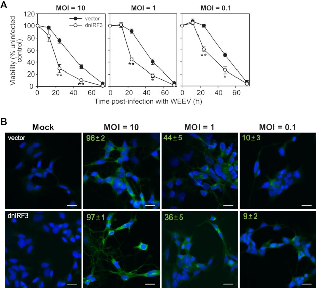
IRF-3 enhances neuronal survival in BE(2)-C/m cells in response to WEEV infection irrespective of viral inoculum. (A) BE(2)-C/m cells stably expressing control vector or dnIRF-3 construct were infected with WEEV at the indicated MOI and percent viability relative to mock-infected controls was measured from 12 to 72 hpi. *, P < 0.05; **, P < 0.005 (compared to WEEV-infected cells expressing control vector at the corresponding time point). (B) Immunofluorescence microscopy images of BE(2)-C/m cells stably expressing control vector (upper images) or dnIRF-3 construct (lower images) infected with WEEV at the indicated MOI and analyzed at 12 hpi. Representative overlaid images from one of two independent experiments are shown, where blue indicates 4′,6-diamidino-2-phenylindole (DAPI)-stained nuclei and green indicates WEEV-infected cells. We quantitated the percentage of infected cells by analyzing four separate fields obtained at ×20 magnification, which corresponded to enumerating ∼250 cells per group. Values in the upper left corners indicate means ± standard deviations (SD) of the percentage of cells positive for WEEV antigens. Scale bars, 25 μm.

