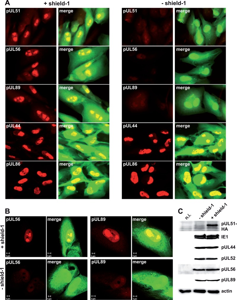
Fig 6.
Expression and localization of the terminase proteins pUL56 and pUL89 in the presence or absence of pUL51. (A) HFF were infected with HCMV-UL51-ddFKBP and cultivated with or without shield-1. On day 4 p.i., cells were examined by immunofluorescence microscopy using the indicated antibodies. EGFP expression marks the infected cells. (B) Cells from the same experiment were also investigated by confocal laser scanning microscopy. (C) Lysates from cells infected as described for panel A were prepared on day 4 p.i. and analyzed by immunoblotting using an anti-HA antibody (for pUL51) or antibodies directed against the indicated proteins. n.i., noninfected cells.

