Fig 4.
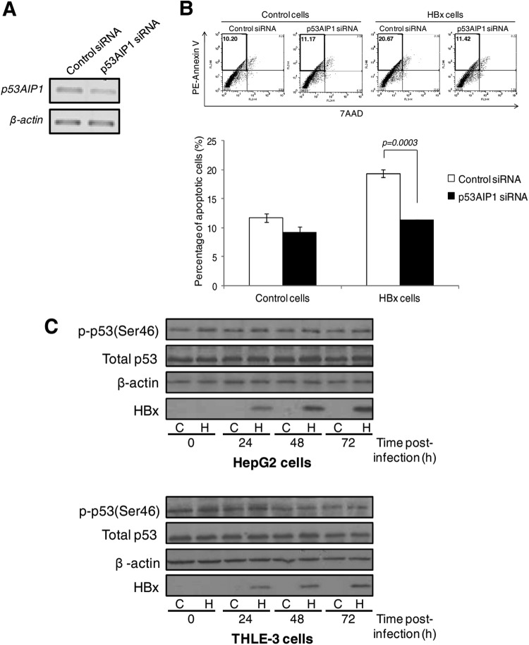
Increased p53AIP1 expression mediates HBx-induced apoptosis. (A) Transient p53AIP1 knockdown using specific siRNA compared to control siRNA measured by qPCR. (B) Apoptosis profiles of p53AIP1-specific or control siRNA-treated control and HBx UV-treated HepG2 cells from a representative set of experiments (top) and summarized from 3 independent experiments shown in percentages (bottom). (Top) The percentage of apoptotic cells in each sample is shown in the upper left quadrant. All error bars show standard errors of the mean from triplicate experiments. (C) Immunoblot of phosphorylated p53 Ser46 levels using a modification-specific antibody in HBx and control HepG2 (top) and THLE-3 (bottom) cells harvested at various time points over 72 h. Immunoblots of total p53, β-actin, and HBx expression are also shown.

