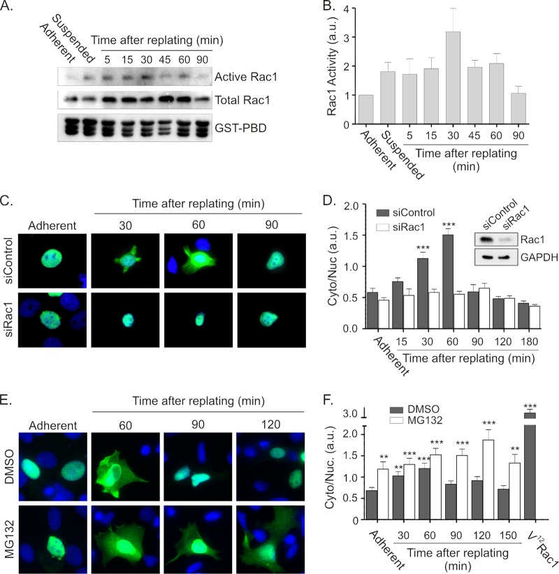
Fig 5.
Cell spreading stimulates Net1A relocalization in a Rac1-dependent and proteasome-regulated manner. (A) MCF7 cells were replated on collagen-coated dishes for different lengths of time and then lysed and tested for endogenous Rac1 activation in GST-PBD assays. Shown are results of a representative experiment. (B) Quantification of Rac1 activation after replating MCF7 cells on collagen. Results shown are the average of three independent experiments. Error bars are standard errors of the means. (C) MCF7 cells were transfected with control of Rac1-specific siRNAs. Two days later, the cells were transfected with an HA-Net1A expression plasmid. Cells were starved overnight and replated on collagen for different lengths of time. Cells were fixed and stained for HA-Net1A (green) and DNA (blue). Results of a representative experiment are shown. (D) Quantification of Net1A subcellular localization. Localization is represented as the ratio of HA-Net1A in the extranuclear space (Cyto) divided by that in the nucleus (Nuc). Error bars are standard errors of the means. Shown in the inset is a representative Western blot, demonstrating Rac1 knockdown. ***, P < 0.001. (E) Proteasome inhibition extends the time of Net1A localization outside the nucleus during cell spreading. MCF7 cells were transfected with an HA-Net1A expression plasmid and then starved and replated on collagen-coated coverslips for different lengths of time in the presence of DMSO or MG132. Cells were then fixed and stained for HA-Net1A (green) and DNA (blue). Shown are results of a representative experiment. (F) Quantification of HA-Net1A localization. Shown are the averages of three independent experiments. Error bars are standard errors of the means. **, P < 0.01; ***, P < 0.001.

