Fig 7.
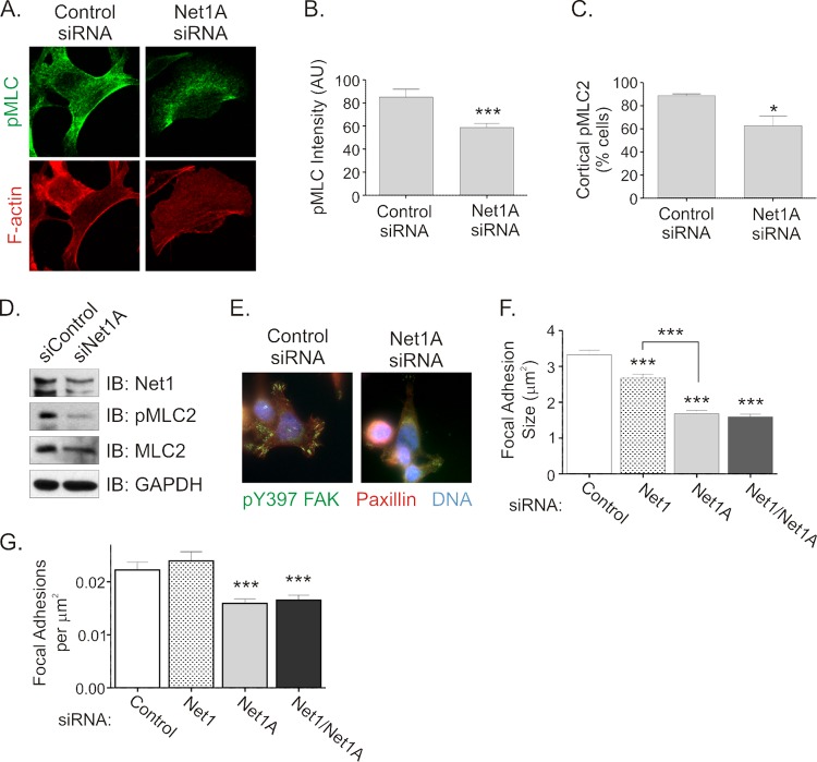
Net1A controls MLC phosphorylation and focal adhesion formation during cell spreading. (A) MCF7 cells were transfected with control or Net1A-specific siRNAs and then replated on collagen-coated coverslips for 1 h. Cells were then fixed and stained for pMLC and F-actin. Shown are representative confocal images. (B) Quantification of pMLC fluorescence intensity. Shown are the averages of three independent experiments. Error bars are standard errors of the means. ***, P < 0.001. (C) Quantification of cortical pMLC staining. Shown are the averages of three independent experiments. Error bars are standard errors of the means. *, P < 0.05. (D) Western blot for pMLC in control or Net1A siRNA-transfected cells. Shown are results of a representative experiment. (E) Net1A knockdown inhibits focal adhesion maturation. MCF7 cells were transfected with control or Net1A-specific siRNAs. Three days later, the cells were replated on collagen-coated coverslips for 1 h. The cells were then fixed and stained for pY397-FAK (green), paxillin (red), and DNA (blue). Shown are representative images. (F) Quantification of focal adhesion size. MCF7 cells were transfected with the siRNAs shown and replated on collagen-coated coverslips for 1 h. Focal adhesions containing pY397-FAK and paxillin were quantified. Error bars are standard errors of the means. ***, P < 0.001. (G) Quantification of focal adhesions per cell area (in μm2). Cells were transfected and processed as for panel F. Phospho-Y397-FAK- and paxillin-positive focal adhesions were quantified, and values were then divided by the areas of the cells. Error bars are standard errors of the means. ***, P < 0.001.

