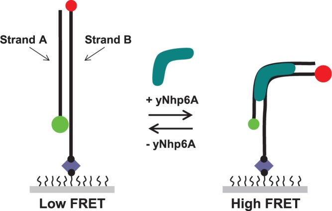
Figure 1.

Schematic design of smFRET experiments. The DNA targets used in this study were designed so that the donor (Cy3, green circle) and acceptor (Cy5, red circle) are attached to opposite termini of the DNA duplex (black lines). Decreases in DNA end-to-end distance induced by yNhp6A (aqua) binding and kinking result in a measurable increase in the efficiency of energy transfer for each individual FRET pair. The DNA-only state is in a ‘low FRET’ state, and the yNhp6A-bound DNA is in a ‘high FRET’ state. Each DNA molecule is immobilized to the surface of a polymer-coated slide through a strong biotin (black circle)–streptavidin (blue diamond) interaction.
