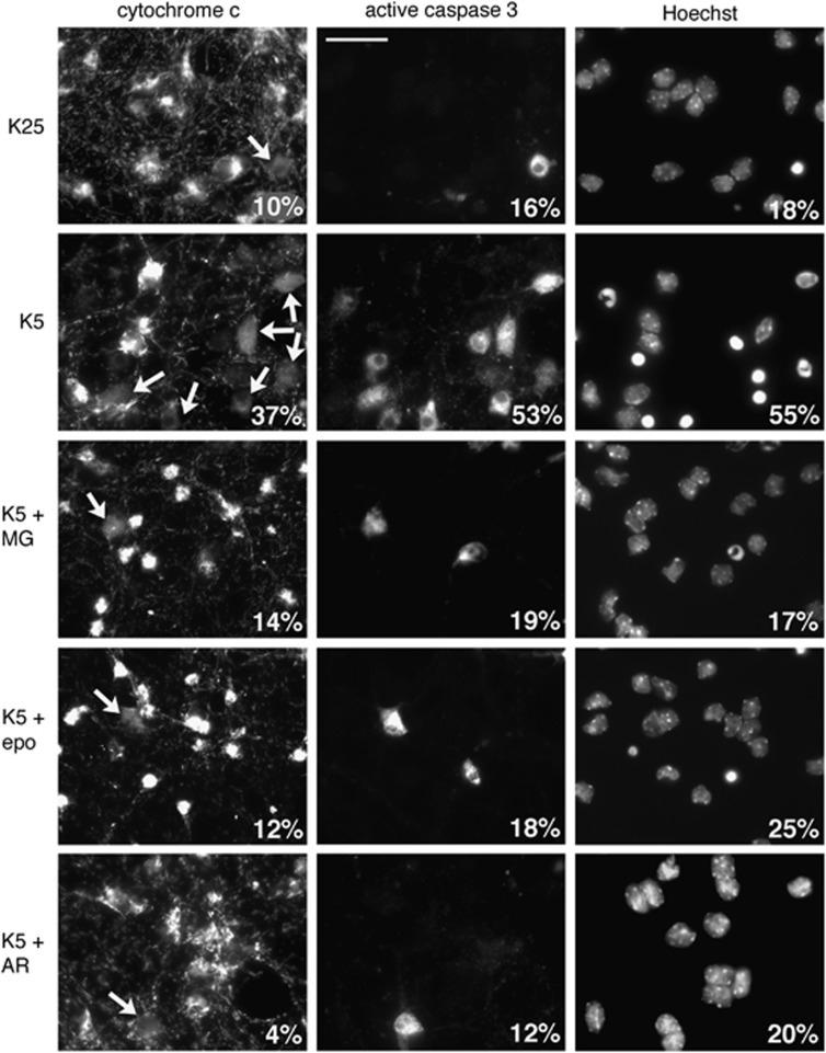
Figure 2.
Proteasome and GSK3 inhibitors protect neurons from apoptosis by preventing cytochrome c release and caspase activation. CGN primary cultures were washed and switched to serum-free medium containing either 25 mM KCl (K25) or 5 mM KCl (K5) in the presence or absence of 20 μM MG-132 (MG), 10 μM epoxomicin (epo) or 10 μM AR-A014418 for 8 h. After fixation, nuclear condensation was visualized by Hoechst 33258 staining. Cytochrome c subcellular localization and caspase 3 activation were detected by immunofluorescence. In healthy neurons, cytochrome c immunostaining is intense and punctate both in cell bodies and in neurites (axons and dendrites), indicating mitochondrial localization. In apoptotic neurons, the staining is faint and diffuse, indicating that cytochrome c has been released from mitochondria. At late stages of apoptosis, the staining disappears because cytochrome c is rapidly degraded after release. The percentages of neurons with a condensed nucleus, showing a diffuse staining for cytochrome c or positive for active caspase 3 are indicated for each condition (500 neurons scored in a blinded manner for each condition). Arrows indicate cell bodies with a diffuse cytochrome c staining. Scale bar is 20 μm. Data are representative of three independent experiments

