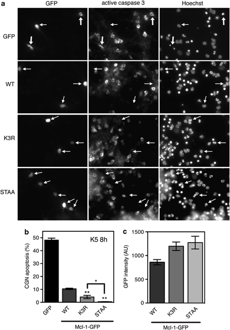
Figure 4.
Neuroprotective effect of the different forms of Mcl-1. (a) CGN primary cultures were transfected with GFP (used as a negative control) or the different forms of Mcl-1 fused to GFP, for 16 h. Then, neurons were incubated in K5 medium for 8 h. Caspase 3 activation was determined by immunofluorescence and nuclear condensation was visualized by Hoechst staining. Thick arrows indicate GFP-expressing neurons that undergo apoptosis, whereas thin arrows indicate healthy neurons expressing GFP or the different forms of Mcl-1-GFP. (b) The percentage of apoptosis among transfected neurons was assessed by examining cell morphology and nuclear condensation of GFP-positive neurons. Data are the mean±S.D. of three independent experiments. **P<0.001 significantly different from apoptosis observed in neurons expressing Mcl-1(WT)-GFP (ANOVA followed by Student-Newman–Keuls multiple comparison test). The difference observed between neurons expressing Mcl-1(K3R)-GFP and Mcl-1(STAA)-GFP is also significant (*P<0.01 using the same statistical tests). (c) GFP intensity (AU: Arbitrary Unit) in the cell body of transfected neurons was measured using MetaMorph. Data are the mean±S.E.M. of about 100 neurons for each condition

