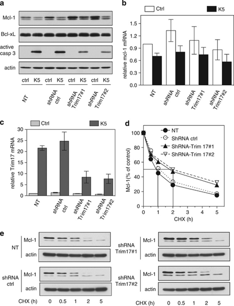
Figure 5.
Silencing of Trim17 favours Mcl-1 stabilization. (a) CGNs were left untreated (non-transduced: NT) or were transduced with lentiviral particles expressing shRNA sequences (one control and two against Trim17) one day after plating. At DIV 6, neurons were incubated for 6 h in K5 medium or maintained in the initial culture medium (ctrl). Then, proteins were analyzed by western blot using antibodies against Mcl-1, Bcl-x, active caspase 3 and actin. (b and c) CGNs were transduced or not as in (a). Then, neurons were incubated for 4 h in K5 medium or maintained in the initial culture medium (ctrl). Total RNA was extracted and the mRNA levels of mcl-1 (b) and Trim17 (c) were estimated under each condition by quantitative RT-PCR. Fold change was calculated by comparison with non-transduced neurons maintained in the initial culture medium. Data are means±s.d. of triplicate measurements of one experiment that is representative of three independent experiments. (d and e) CGNs were transduced or not as in (a) and incubated for increasing times with 10 μg/ml cycloheximide (CHX). Proteins were analyzed by western blot using antibodies against Mcl-1 and actin. (d) The intensity of the Mcl-1 bands presented in a representative experiment performed as in (e) was estimated and expressed as a percentage of the corresponding value for time zero

