Figure 4.
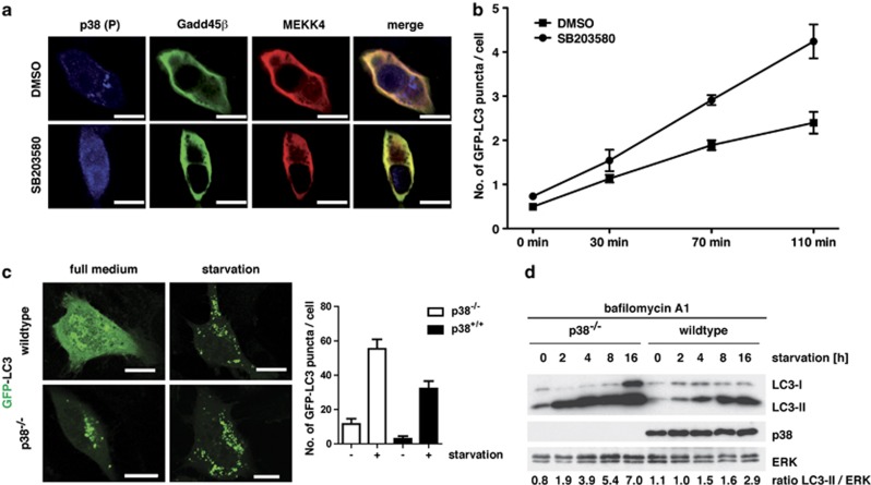
Lack of active p38 accelerates autophagy. (a) NIH/3T3 cells were transiently transfected with V5-tagged Gadd45β and HA-tagged MEKK4. At 24 h post transfection, the cells were fixed and stained for endogenous phosphorylated p38 (blue), HA-tagged MEKK4 (red) and the V5-tagged Gadd45β (green). In addition, the cells were treated with 500 nM of the p38 inhibitor SB203580 or DMSO as solvent control. Bars, 10 μm. (b) GFP-LC3 expressing NIH/3T3 cells were cultured in the presence or absence of 500 nM SB203580 in HBSS and followed by life cell imaging for the indicated time points. Autophagosomes were counted in 40 cells and the mean number of autophagosomes per cell was calculated from two independent visual fields. Error bars represent S.E.M. (c) WT and p38−/− MEFs were transfected with GFP-LC3 as a marker for autophagy. At 24 h post transfection, cells were cultured for 4 h in HBSS medium to induce autophagy. Subsequently, samples were fixed and analyzed by confocal microscopy (left panel). Bars, 10 μm. The number of autophagosomes was counted in 40 cells per condition (right panel). (d) WT and p38−/− MEFs were cultured for up to 16 h in HBSS medium to induce autophagy. In order to prevent LC3-II degradation, cells were pretreated with 100 nM bafilomycin A1 for 1 h. Subsequently, cellular lysates were prepared and analyzed by immunoblotting using the indicated antibodies. Densitometric quantification of the LC3-II to ERK ratio is depicted at the bottom

