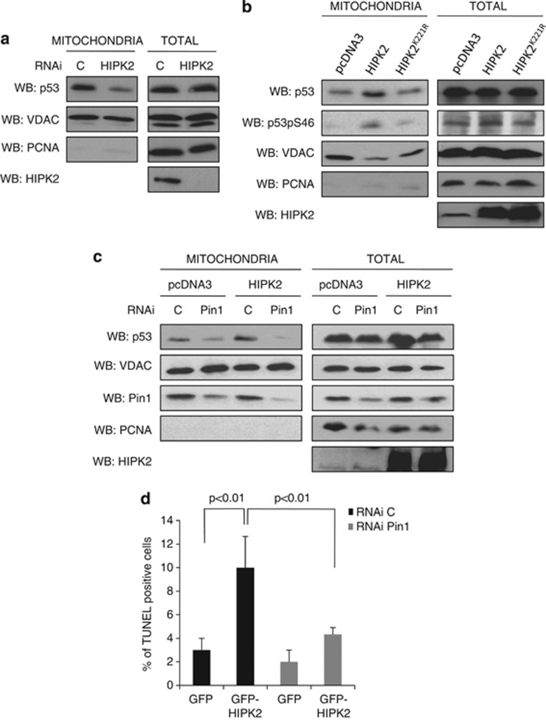
Figure 4.
HIPK2 and Pin1 cooperate to induce p53-dependent direct apoptosis. (a) HIPK2 promotes mitochondrial localization of p53. Accumulation of endogenous p53 within the mitochondrial fraction of HCT116 p53+/+ cells was analyzed by WB upon RNAi-mediated knockdown of HIPK2 and treatment with doxorubicin 1 μM for 6 h. C: control RNAi. (b) Mitochondrial localization of endogenous p53 upon overexpression of either wild-type HIPK2 or the catalytically inactive mutant HIPK2K221R in HCT116 p53+/+ cells and treatment with 1 μM doxorubicin for 6 h was analyzed as in a. (c) Pin1 is required for the ability of HIPK2 to promote mitochondrial localization of p53. The effect of wild-type HIPK2 overexpression on the mitochondrial localization of endogenous p53 was evaluated as in a in HCT116 p53+/+ cells treated with doxorubicin 1 μM for 6 h upon RNAi-mediated knockdown of Pin1 expression. (d) To estimate the roles of HIPK2 and Pin1 in inducing transcription-independent apoptosis, HCT116 p53+/+ cells transfected with the indicated combinations of GFP-HIPK2 expression vector and Pin1-specific RNAi were treated with α-amanitin 10 μg/ml. Apoptosis of GFP-expressing cells was then evaluated by TUNEL assays. The graph shows the mean results and S.D. of three independent experiments

