FIG. 1.
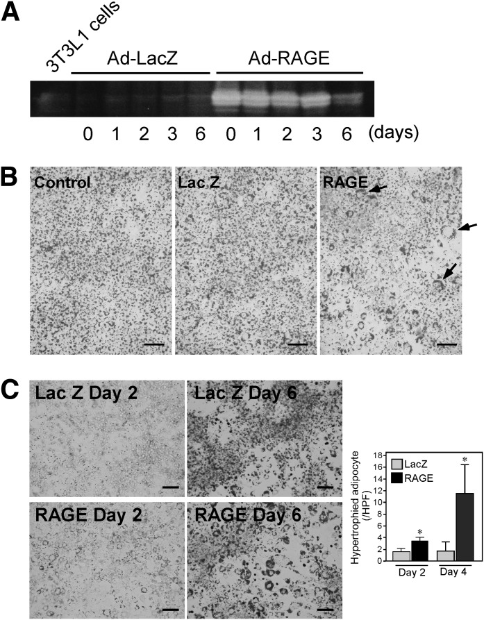
RAGE overexpression in 3T3L1 preadipocytes accelerates adipocyte hypertrophy. Confluent 3T3L1 cells were infected with human RAGE- or control β-galactosidase (LacZ)–expressing adenovirus. At day 0, differentiation was initiated by the addition of 10% FBS, 1 μmol/L insulin, and 0.5 mmol/L isobutylmethyl xanthine, and 0.25 μmol/L dexamethasone for 2 days. Then, the cells were cultured for 2 more days in 10% FBS and 1 μmol/L insulin (day 2–4). The cells were then maintained in 10% FBS medium for 2 days (day 4–6). A: At indicated days of differentiation, RAGE protein expression in 3T3L1 cells was determined by Western blot analyses using goat anti-human RAGE antibody. Adenoviral overexpression of RAGE was maintained up until day 6 during adipocyte differentiation. B: Oil Red O staining of 3T3-L1 adipocytes was performed to determine adipogenesis at day 6. More hypertrophic adipocytes with ring-like lipid staining (arrows) were observed in RAGE-overexpressed cells. Control represents 3T3L1 cells without adenoviral infection. C: Oil Red O staining was performed at days 2 or 6 during differentiation. Numbers of hypertrophic adipocytes, as defined as cells with ring-like lipid droplets, were counted in five random fields (×20 power) scanned by BioZero fluorescent microscope (Keyence). Each column represents mean ± SD. Scale bars, 100 μm. *P < 0.05, Student t test. HPF, high-power field.

