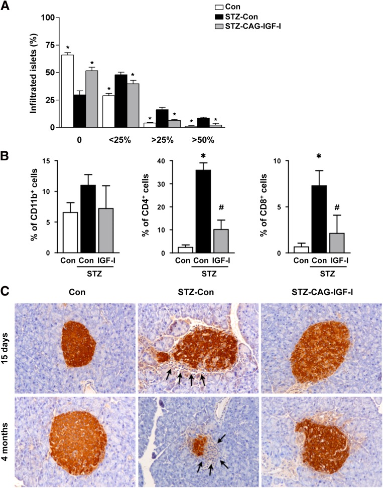
FIG. 3.
Decreased pancreatic infiltration following IGF-I treatment. A: Insulitis score was determined in control (Con), STZ-Con, and STZ-CAG-IGF-I animals 15 days after STZ treatment. No infiltration (0), peri-insulitis (<25%), moderate insulitis (<50%), and severe insulitis (>50%). Results are mean ± SEM of four mice per group. *P < 0.05 vs. STZ-Con. B: Percentage of infiltrating macrophages (CD11b+), T CD4+ cells, and T CD8+ cells in islets. The presence of these cells in islets was quantified by flow cytometry from pancreas extracts 15 days after STZ treatment. Results are mean ± SEM of three animals per group. *P < 0.05 vs. Con. #P < 0.05 vs. STZ-Con. C: Immunohistochemical detection of insulin in pancreatic sections of Con, STZ-Con, and STZ-CAG-IGF-I–treated mice at 2 weeks and 4 months after STZ treatment. Arrows indicate lymphocytic infiltrate in islets. Data are representative of at least three independent experiments. (A high-quality digital representation of this figure is available in the online issue.)

