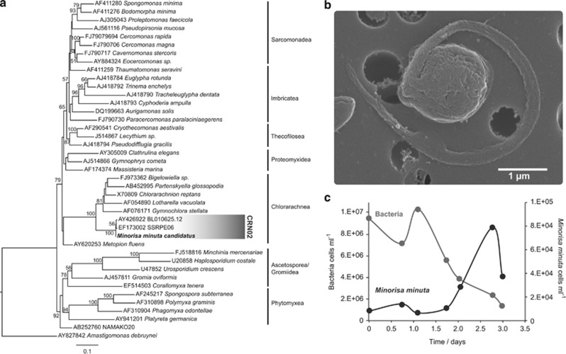
Figure 1.
(a) ML phylogenetic tree with complete 18S rDNA sequences showing the position of Minorisa minuta within the Cercozoa. The scale bar indicates 0.1 substitutions per position. The coverage of the specific probe CRN02 is shown in grey. (b) SEM image of a Minorisa minuta cell of 1.6 μm in size and possessing a single flagellum. (c) Growth of Minorisa minuta with natural bacteria as prey in a batch culture.

