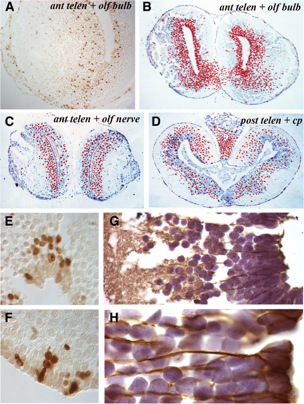
Figure 4.
Migration patterns and glial fibrillary acidic protein expression in the telencephalon. (A) Single section through the anterior telencephalon and olfactory bulb taken 4 weeks after overnight BrdU labeling shows a stream of cells moving laterally into the olfactory bulb. (B) Cumulative positions of BrdU+ cells in the anterior telencephalon and olfactory bulb level 4 weeks after overnight labeling. (C) Cumulative positions of BrdU+ cells in the anterior telencephalon and olfactory nerve level 4 weeks after overnight labeling showing a decreased number of BrdU+ cells in the VZ. (D) Cumulative positions of BrdU+ cells in the posterior telencephalon 4 weeks after overnight labeling showing a decreased number of BrdU+ cells in the VZ. (E,F) Patterns of BrdU+ cells in and adjacent to the VZ 3 weeks after overnight labeling. (G,H) Glial fibrillary acidic protein immunocytochemistry of the normal telencephalon at low power (G) and high power (H) to show the cytoplasmic extensions of radial glial cells. BrdU: bromodeoxyuridine; VZ: ventricular zone.

