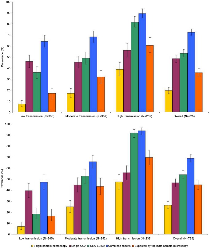
Figure 2. Prevalence of intestinal schistosomiasis in preschoolers at baseline and follow-up.
Microscopy was conducted on duplicate Kato-Katz thick smears from the same stool sample; a single CCA tests was conducted on a single urine sample; SEA-ELISAs were conducted in the field; ‘combined results’: if positive for any test then considered child as positive; and mathematical model by Jordan and colleagues (‘Triplicate stool samples microscopy’) [29] Baseline results represented in top section, and follow-up results represented in bottom section.

