
Figure 3.
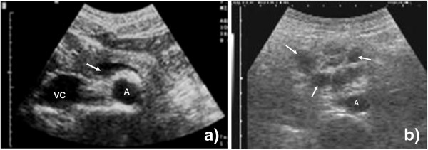
Probe position 1b. (a) Vascular structures (abdominal aorta (A), inferior vena cava (VC), splenic vein (arrow)) of the upper abdomen are visible. (b) Multiple round hypoechoic structures are visible (arrow). These represent pathologically enlarged lymph nodes close to the aorta (A).