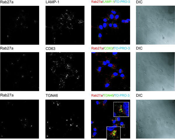
Figure 2.
Subcellular localization of Rab27a in HOG cells. A. HOG cells cultured in DM were fixed and processed for confocal double-label indirect immunofluorescence analysis with anti-Rab27a polyclonal antibody and antibodies against LAMP-1, CD63 and TGN-46. Primary antibodies were detected using Alexa Fluor 555 and 488 secondary antibodies. Images correspond to the projection of the planes obtained by confocal microscopy. Colocalization (yellow spots) was detected between Rab27a and TGN-46. The squares show enlarged images corresponding to a confocal slice of 0.8 μm. (DIC: Differential Interference Contrast).

