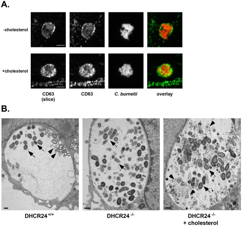
Figure 6. The lumenal contents of the C. burnetii vacuole are altered in the absence of cholesterol.
(A). Fluorescence micrographs of C. burnetii-infected −cholesterol and +cholesterol MEFs stained for the late endosome/multivesicular body marker CD63 (green) and C. burnetii (red) at 4 dpi. Unlike +cholesterol MEFs, CD63 was not found in the lumen of vacuoles within −cholesterol MEFs. The first panel shows a single confocal slice through the middle of the vacuole, while the second panel is a maximum intensity Z projection of the entire vacuole. Scale bars = 5 µm. (B). Transmission electron microscopy revealed multilamellar membranous structures (arrowheads) within vacuoles harboring C. burnetii (arrows) in infected DHCR24+/+ and DHCR24−/− +cholesterol MEFs, but not DHCR24−/− −cholesterol MEFs. Scale bars = 500 nm.

