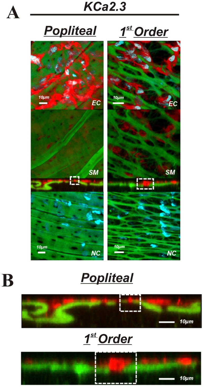
Figure 5. KCa2.3 in skeletal muscle arteries.
A: Skeletal muscle popliteal (left) and 1st order (right) arteries immunolabeled for KCa2.3 (top 3 panels) or no primary antibody control (bottom panel). The top image represents a composite of DAPI-stained nuclei in the endothelium, autofluorescence of the internal elastic lamina (green), and KCa2.3 immunolabeling (red), and a compressed z-stack image endothelial cell surface (EC, top panel). The second image is a compressed z-stack image from the perspective of the smooth muscle face (SM), the third image is a cross-sectional slice, and the fourth is a compressed z-stack image of a no primary antibody control (NC). Red staining indicates endothelial expression of KCa2.3. B: Magnified image of the cross-sectional view of the IEL. Note the sections highlighted by dotted square boxes indicating larger MEC and area of KCa2.3 staining in 1st order arterioles. Bar = 10 µm. Representative of images from tissue isolated from at least 3 animals.

