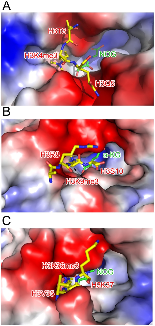
Figure 7. Comparison of the substrate peptide binding modes of c-JMJ703 and c-JMJD2A.
(A) Binding of H3K4me3 peptide by c-JMJ703. (B) Binding of H3K9me3 by c-JMJD2A. (C) Binding of H3K36me3 by c-JMJD2A. c-JMJ703 and c-JMJD2A are shown as electrostatic surface and the substrate peptides are shown as yellow sticks and the residues are labeled in red. NOG and α-KG are shown as green sticks and labeled.

