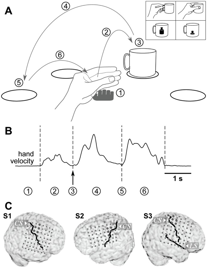
Figure 1. Experimental paradigm.
A: Task layout. The subject’s hand was resting palm down (1) on a central spot, marked by the grey hand pictograph. A reaching movement (2) was initiated by the subject (self-paced) and a cup was grasped (3) at one of four marked positions (circles) and carried (4) to one of the remaining three positions (self-chosen). There, the cup was released (5) and the hand returned (6) to the central resting position. Grasps of the object varied with respect to the applied grasp type (precision or whole-hand grip, self-chosen in every trial) and object weight (switched between two cups every 15–16 trials), pictured in the upper right inset. B: Sample profile of hand velocity during one trial. Actions labeled by numbers in (a) are marked at their respective times. C: Electrode implantations in all three subjects. The position of the central sulcus and the lateral sulcus (S3 only) relative to the electrodes are shown by black lines. Electrode contacts of hand-arm motor cortex are marked by a black circle with a black dot in the center.

