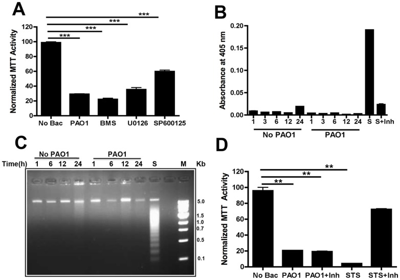
Figure 6. Toxicity of PAO1 toward RAW 264.7 cells.
A) RAW 264.7 cells were exposed to PAO1 in the presence of different kinase inhibitors: SP600125 (for JNK, 10 mg/ml), U0126 (for MEK, 10 mg/ml) and BMS345541 (for IKK, 10 mg/ml). MTT assays quantitated cell viability after 1 day. B) RAW 264.7 cells were treated with PAO1 (MOI 10, 1 h) and washed. Caspase-3 activity was measured by colorimetric assays after 1–24 h. Control cells not exposed to bacteria were collected at the same time points. Sample treated with staurosporine (STS, 1 µg/ml) with or without caspase-3 inhibitor (Inh) for 3 h were used as a positive control. C) RAW 264.7 cells were exposed to PAO1 (MOI 10) for 1 h. DNA was isolated from cells at the indicated time points and analyzed by agarose gel electrophoresis. Control cells were not exposed to PAO1. Cells incubated with staurosporine (S) for 3 h were used as a positive control. M denotes the DNA ladder. D) RAW 264.7 cells were exposed to PAO1 +/− prior incubation with caspase 3 inhibitor (Inh). The viability of the cells was measured by MTT assay after 2 days. Cells treated with staurosporine were used as a positive control. Data are indicative of three repeat experiments with similar results and error bars represent standard deviations. (***) designates p<0.0003.

