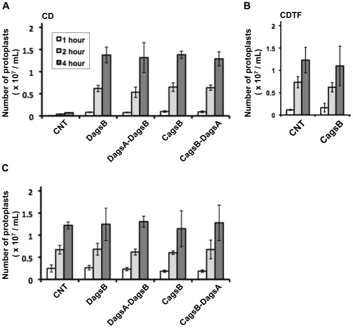
Figure 6. Cell wall susceptibility of the control (CNT), agsB disruption (DagsB), agsA agsB double disruption (DagsA-DagsB), CagsB, and CagsB with agsA disruption (CagsB-DagsA) strains to Lysing Enzymes.
(A) Susceptibility of mycelia cultured in CD liquid medium (agsB-repressing conditions). Mycelia cultured in CD medium for 24 h (30 mg fresh weight) were digested in reaction buffer (10 mM phosphate buffer, pH 6.0) containing 10 mg/mL Lysing Enzymes. After 1, 2, and 4 h of incubation at 30°C, the number of protoplasts in each sample was determined using a hemocytometer. Error bars represent the standard deviations (n = 3). All values of the mutants differed significantly from that of the control strain. (B) Susceptibility of the control and CagsB strains cultured in CDTF liquid medium (agsB-inducing conditions). Mycelia cultured in CDTF medium for 24 h (30 mg fresh weight) were digested, and the number of protoplasts in each sample was determined according to the method described above. None of the differences were statistically significant. (C) Effect of combined treatment with α-1,3-glucanase and Lysing Enzymes on protoplast formation from fungal mycelia. Mycelia cultured in CD medium for 24 h were digested in reaction buffer containing both Lysing Enzymes and α-1,3-glucanase. The number of protoplasts generated in each sample was determined according to the method described above. None of the differences were statistically significant.

