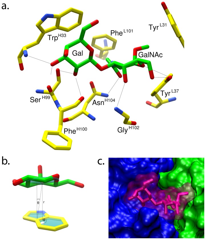
Figure 3. The binding interactions predicted from the docked model of the TF-disaccharide bound to JAA-F11.
a. Validated model of the bound minimal determinant (green carbon frame) in the mAb binding site, hydrogen atoms removed for clarity. Protein residues (yellow carbon frame) involved in hydrogen bonds (black lines) or hydrophobic interactions with the TF antigen are shown. b. Depiction of the stacking interaction between the Gal and the TrpH33 (r = 4.0 Å, θ = 12.1°, φ = 90.3°); the geometry of this interaction is comparable to literature values[53] (lower left). c. Solvent accessible surface of the bound minimal determinant (magenta) and the Fab (showing VH and VL regions in blue and green, respectively.

