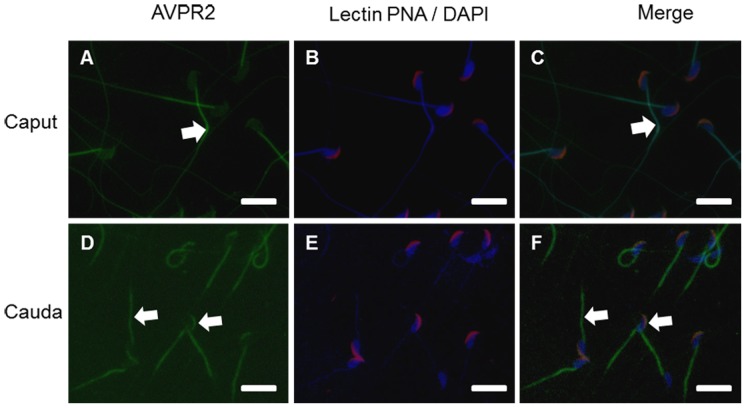
Figure 1. Localization of AVPR2 in epididymal mouse spermatozoa.
Immunohistochemistry staining of mouse caput (A, B, C) and cauda (D, E, F) spermatozoa. B and E are merged images of acrosome (Lectin PNA conjugated Alex 647, red) and nucleus (DAPI, blue). C and F are merged image of AVPR2, acrosome and nucleus. Arrows directed AVPR2 (A, C, D, and F). Images obtained using Nikon TS-1000 microscope with NIS Elements image software (Nikon, Japan). Bar = 10 µm.

