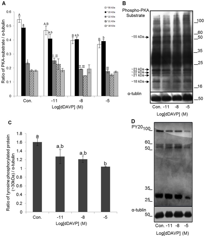
Figure 5. Effect of dDAVP on PKA activity and protein tyrosine phosphorylation.
(A) Density of PKA substrates in various treatments (Open Bar: ∼55 kDa, Black Bar: ∼23 kDa, Grey Bar: ∼22 kDa, Stripe Bar: ∼21 kDa, and Dot Bar: ∼18 kDa). Data represent mean ± SEM, n = 3. Values with different superscripts (A,B,a,b,I,II,III) were significantly different between control and treatment groups by One-way ANOVA (p<0.05). (B) Phospho-PKA substrates were probed with anti-phospho-PKA substrates; lane 1: Control, lane 2: 10−11 M dDAVP, lane 3: 10−8 M dDAVP, lane 4: 10−5 M dDAVP. (C) Density of tyrosine phosphorylated protein (∼30 kDa) in various treatments. Data represent mean ± SEM, n = 3. Values with different superscripts (a,b) were significantly different between control and treatment groups by One-way ANOVA (p<0.05). (D) tyrosine phosphorylated proteins were probed with PY 20; lane 1: Control, lane 2: 10−11 M dDAVP, lane 3: 10−8 M dDAVP, lane 4: 10−5 M dDAVP.

