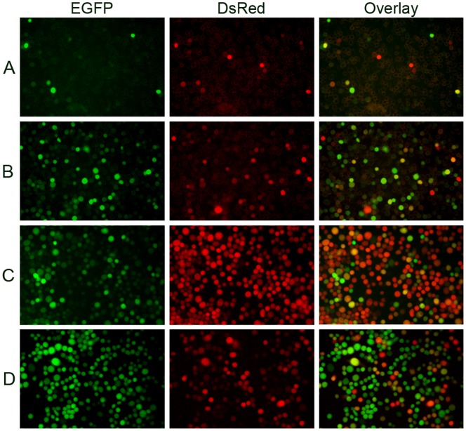
Figure 4. Microscopic analysis of Sf9 cells co-infected with vAcBacGFP and vAcBacDsRed.
Sf9 cells were simultaneously infected with vAcBacGFP and vAcBacDsRed viruses. Protein expression was observed 48 hpi. Many cells were found co-expressing red and green fluorescent proteins even at low multiplicity. A. Cells were infected with 0.05 MOI of each virus. B. Cells were infected with 0.5 MOI of each virus. C. Cells were infected with vAcBacGFP and vAcBacDsRed at a ratio of 1∶4 respectively. D. Cells were infected with vAcBacGFP and vAcBacDsRed at the ratio of 4∶1 respectively.

