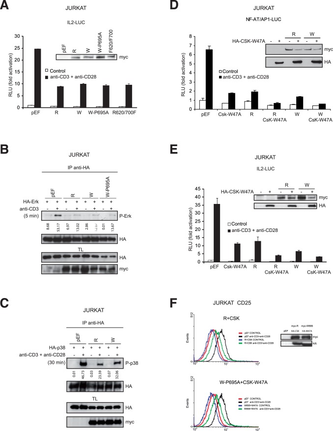
Figure 4. LYP/CSK interaction is not required to regulate TCR signaling.
A, Activation of a luciferase reporter gene driven by the IL-2 minimal promoter in Jurkat cells co-transfected with different myc-LYP plasmids, as indicated. The insert shows the expression of the LYP proteins as detected by IB. B, Erk activation was assayed in Jurkat cells transfected with different versions of LYP, as indicated, and stimulated with anti-CD3 Ab for 5 min. Erk was immunoprecipitated from lysates of these cells and its phosphorylation was detected by IB. Expression was verified in total lysates (TL) by IB. Phospho-ERK (P-Erk) blot was measured by densitometry and the data were expressed as arbitrary units under the blot.C, As in B, p38 activity was evaluated in Jurkat T cells stimulated with anti-CD3 and anti-CD28 Ab for 30 min by IP of HA-p38 and IB with a specific antibody for dually phosphorylated p38. Phospho-p38 (P-p38) blot was measured by densitometry scanning and the data were expressed as arbitrary units under the blot. D, Activation of a luciferase reporter gene driven by the NF-AT/AP1 site of IL-2 promoter in Jurkat cells co-transfected with LYPR, LYPTW, and CSK-W47A plasmids, as indicated. Expression of LYP and CSK proteins as detected by IB is shown in the insert. E, Activation of a luciferase reporter gene driven by the IL-2 minimal promoter in Jurkat cells transfected with LYPR, LYPW, and CSK-W47A plasmids. The insert shows the IB of LYP and CSK proteins. R, LYPR; W, LYPW. F, Expression of CD25 in Jurkat cells transfected with the plasmids indicated was measured by flow cytometry upon stimulation with anti-CD3 plus anti-CD28 antibodies for 24 hours. Expression of LYP and CSK proteins as detected by IB is shown in the insert.

