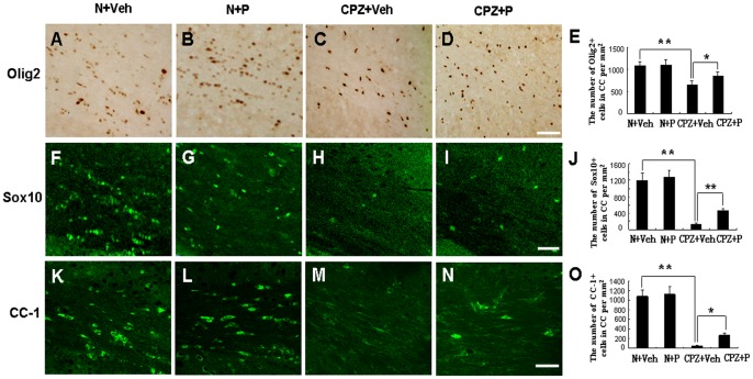
Figure 5. Progesterone rescued the loss of oligodendroglial cells in the cuprizone-induced mice.
Olig2-positive cells were observed in the corpus callosum of N+Veh (A), N+P (B), CPZ+Veh (C), and CPZ+P group (D). (E) Statistical analyses showed that Olig2-positive cells increased in the CPZ+P group compared with the CPZ+Veh group. Bar = 50 µm. *CPZ+P vs. CPZ+Veh group, P<0.05; **CPZ+Veh vs. N+Veh group, P<0.01. Sox10-positive cells were observed in the corpus callosum of mice in N+Veh (F), N+P (G), CPZ+Veh (H), and CPZ+P group (I). (J) Statistical analyses showed that the number of Sox10-positive cells increased in the CPZ+P group compared with the CPZ+Veh group. Bar = 50 µm. **CPZ+Veh vs. N+Veh, CPZ+ P vs. CPZ+Veh, P<0.01. CC-1+ mature oligodendrocytes were observed in the corpus callosum of mice in N+Veh group (K), N+P group (L), CPZ+Veh group (M), and CPZ+P group (N). (O) Statistical analysis showed that CC-1-positive cells increased in the CPZ+P group compared with the CPZ+Veh group. Bar = 50 µm. *CPZ+P vs. CPZ+Veh group, P<0.05; **CPZ+Veh vs. N+Veh group, P<0.01.

