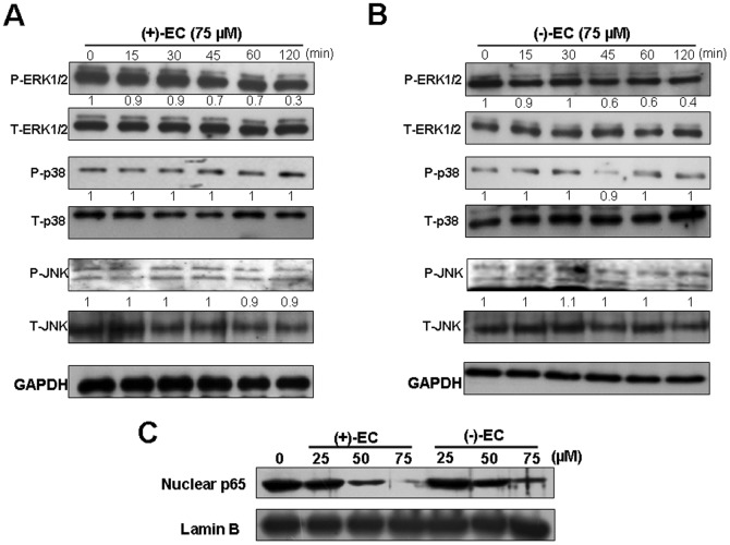
Figure 4. Inhibitory effect of the EC isomers on HCV-induced ERK and NF-κB pathways.
Ava5 cells were treated with (+)-EC (A) and (−)-EC (B) at a concentration of 75 µM for indicated times (0, 15, 30, 45, 60, 120 min). Total cell lysates were prepared for Western blot analysis with specific antibody against P-ERK1/2, T-ERK, P-p38, T-p38, P-JNK, T-JNK, and GAPDH (loading control). (C) Ava5 cells were treated with the indicated concentrations of (+)-EC and (−)-EC for 3 days. The nuclear extracts were prepared for Western blot analysis with anti-p65 and anti-Lamin B. The relative blot intensities were quantified by densitometric scanning. The densitometry values were normalized to GAPDH and untreated values set as 1. Each experiment was performed in triplicate.

