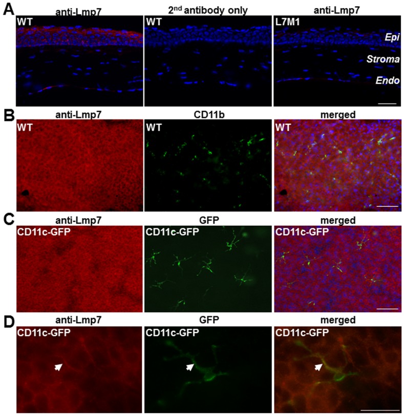
Figure 1. Expression of immunoproteasome in murine corneal epithelium.
( A ) Immunostaining with anti-Lmp7 antibody in WT corneal sections showed expression of immunoproteasome. Negative controls included staining with secondary antibody only (WT) and staining with anti-Lmp7 in immunoproteasome knock-out mice (L7M1). Red: Lmp7; Blue: DAPI-stained nuclei (100× magnification; scale bar, 50 µ). “Epi”: epithelium; “Endo”: endothelium. ( B ) Immune reactions of whole-mount corneas from WT mice using anti-Lmp7 (red) and anti-CD11b (green) antibodies. Images were taken at different focal depths of the same visual field in the peripheral cornea. Merged image shows anti-Lmp7 staining was not limited to the myeloid cells. Blue: DAPI-stained nuclei (100× magnification; scale bar: 150 µ). ( C ) Whole-mount corneas from CD11c-DTR-GFP transgenic mice stained with anti-Lmp7 antibody (red). GFP (green) fluorescence was associated with dendritic cells. Merged image showed minimal contribution of dendritic cells to the anti-Lmp7 staining. Blue: DAPI-stained nuclei (100× magnification; scale bar, 150 µ). ( D ) Higher magnification images of CD11c-GFP positive dendritic cells. Arrow heads indicate co-localization of Lmp7 and CD11c signals (merged). Images were taken at 400× magnification; scale bar, 50 µ.

