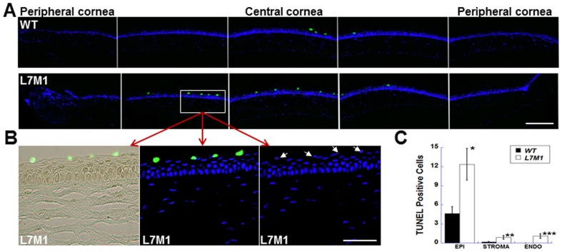
Figure 4. TUNEL staining revealed more apoptosis in uninjured L7M1 corneas.
( A ) Images of corneal sections stained by TUNEL indicated more apoptotic cells in the epithelium of L7M1 mice than of WT mice (Green: TUNEL-positive cells; 25× magnification; scale bar, 150 µ). ( B ) The enlarged TUNEL/bright field (left panel), TUNEL/DAPI (middle panel) and DAPI (right panel) images of the boxed region from L7M1 cornea in (A). The corresponding nuclei of TUNEL-positive cells are indicated by the arrows. (Scale bar, 50 µ) ( C ) Summary of TUNEL-positive cells for each corneal layer. Data are mean ± SEM (n = 10 WT; n = 12 L7M1). *p = 0.014; **p = 0.043; ***p = 0.017.

