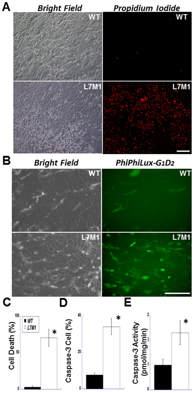
Figure 5. More cell death and higher caspase-3 activity observed in explant-cultured corneal epithelial cells from L7M1 mice.

( A ) Explant-cultured corneal epithelial cells were stained with propidium iodide on day 7 (right panels, 100× magnification; scale bar, 150 µ). The bright field images of the same viewing field are shown in the left panels. ( B ) After 5–6 days of explant culture, WT and L7M1 corneal epithelial cells were stained with a caspase-3 fluorogenic substrate, PhiPhiLux-G1D2 (Green; FITC; 200× magnification; scale bar, 100 µ). ( C ) Summary of the percentage of the PI-positive stained cells in WT and L7M1 cultures. Data are mean ± SEM (n = 11), *p<0.0001. ( D ) Summary of the percentage of cells stained positive for cleaved PhiPhiLux-G1D2. n = 5 WT; n = 9 L7M1; *p = 0.009. ( E ) Summary of caspase-3 activity determined from the cleavage of AC-DMQD-AMC in cell lysates of WT and L7M1 corneal epithelial cells. n = 6 WT; n = 4 L7M1; *p = 0.027.
