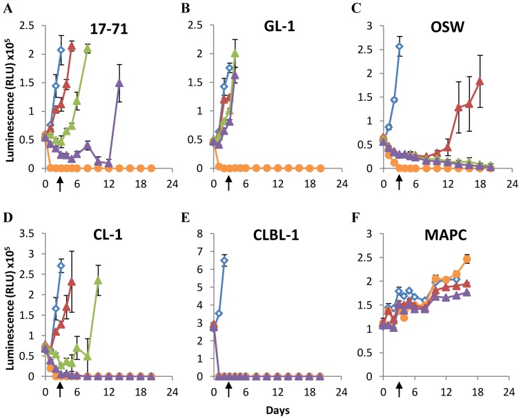
Figure 5. Comparison of Arginase and Asparaginase supplementation on the survival of canine lymphoid cells.
Canine lymphoid cells (A–E), or MAPC (F) were cultured in normal RPMI for 3 days with or without arginase or asparaginase supplementation as indicated below. Enzymes were quenched on day 3 as indicated by the arrows. Cell viability was determined by ATP quantification. Blue diamonds = normal RPMI, red triangles = +0.3 U/ml Asparaginase, green triangles = +1 U/ml Asparaginase, purple triangles = +10 U/ml Asparaginase, Orange circles = +10 U/ml Arginase. Graphs show a representative experiment from three independent experiments with similar results. Error bars represent SD.

