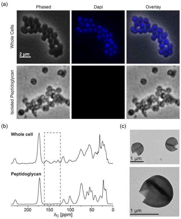
Figure 3. Verification of the purity of the isolated peptidoglycan.
(a) Light microscopy images of DAPI-stained whole cells (top) and isolated peptidoglycan (bottom) from stationary phase cells. The absence of DAPI fluorescence in isolated peptidoglycan indicates that there is no whole-cell contamination. (b) 13C CPMAS spectra of natural abundance whole cells and isolated peptidoglycan. Highlighted in box is the absence of purines in the isolated peptidoglycan spectrum which also confirms that no whole cells remained in the sample. (c) Negative staining TEM images of isolated peptidoglycan.

