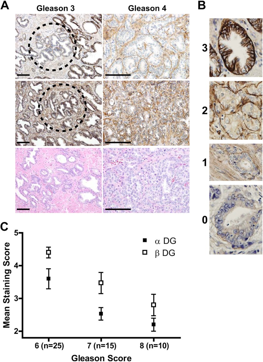
FIGURE 1.
αDG glycosylation and βDG expression are reduced in prostate adenocarcinoma. A, representative images of αDG and βDG immunostained human prostatic tissue showing loss of DG correlating with cancer progression. Scale bar, 100 μm. B, grading was determined using 0–3 grading scale where 0 = basal staining absent; 1 = discontinuous thin basal staining; 2 = thin basal staining with extension between cells or thin dark basal stain without basal-lateral staining; 3 = thick, dark basal staining with basal-lateral staining, characteristic of benign glands. C, mean DG staining of human prostate cancer tissue samples shows that DG expression is reduced across Gleason scores 6–8 (αDG p < 0.0022 and βDG p < 0.0003), while the magnitude of loss of αDG is greater than loss of βDG immunoreactivity.

