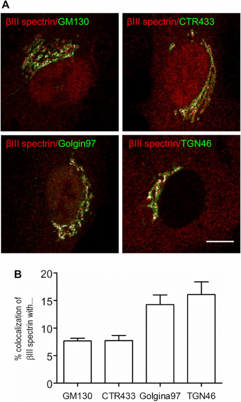
FIGURE 2.
βIII spectrin is enriched in distal Golgi compartments. A, co-localization of βIII spectrin with Golgi markers. RPE1 cells were co-stained with anti-βIII spectrin antibody 33 and antibodies against cis- (GM130), medial- (CTR433), trans-Golgi (Golgin97), or the TGN (TGN46) proteins. Merge pixels are shown in white to facilitate the visualization of the co-labeling. Bar, 10 μm. B, quantitative analysis of the co-localization shown in A.

