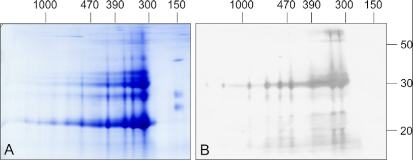
FIGURE 3.
Immunoblotting analyses of a B. napus PSV fraction separated by two-dimensional BN/SDS-PAGE reveals high enrichment of cruciferin. Gels were either stained by Coomassie colloidal (A) or blotted onto nitrocellulose (B). Western blots were incubated with antibodies directed against the α chain of cruciferin. Molecular masses of standard proteins are shown above and to the right of the gel (in kDa).

