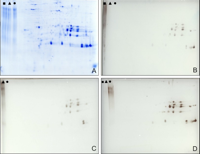
FIGURE 7.
Immunological analysis of cruciferin phosphorylation. Protein storage vacuole fractions were resolved by two-dimensional IEF/SDS-PAGE and blotted onto membranes, and blots were developed using antibodies directed against three different phosphorylation sites. As a control, phosphorylated BSA (phosphorylated at tyrosine (■); phosphorylated at threonine (▴); phosphorylated at serine (●)) was separated within the same gel. A, Coomassie-stained reference gel. B, immunoblot developed with an IgG directed against phosphorylated tyrosine; C, immunoblot developed with an IgG directed against phosphorylated threonine; D, immunoblot developed with an IgG directed against phosphorylated serine. Note that the phosphotyrosine-BSA control on C is not indicated because it partially was beyond the limit of the blot. However, the specificity of the IgG was verified by independent experiments (not shown).

