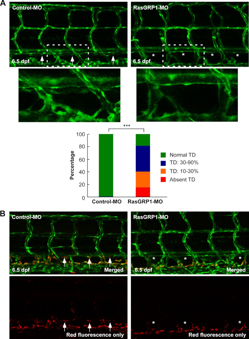
FIGURE 3.
Knockdown of rasgrp1 leads to defects in thoracic duct formation. A, representative confocal images of 6.5 dpf in Fli1:eGFPy zebrafish embryos injected with control MO or RasGRP1-MO. Normal TD formation in the control embryos is marked by arrows, and the absence of the TD in RasGRP1-MO-injected embryos is marked by asterisks. The insets are magnified pictures of the dotted-line boxes. TD formation was quantitatively analyzed by measuring its presence from segments of somite 5 to somite 15, and the result is shown in the bottom panel (n = 120 for control embryos, and n = 187 for RasGRP1 morphants). ***, p < 0.001 by Chi square analysis. B, lymphatic dye uptake assay in Fli1:eGFPy embryos. Alexa Fluor 568 dextran (red) was injected into zebrafish embryos at 6 dpf, and the uptake of the dye was found in control embryos (arrows) but not in RasGRP1-MO-injected embryos (asterisks).

