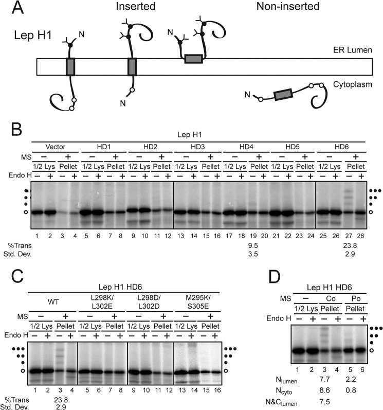
FIGURE 3.
HD6 inserts into RER-derived microsomes in multiple orientations. A, diagram of model Lep H1 used to determine autonomous membrane insertion of individual domains. B and C, HD1–6 domains were analyzed as described in Fig. 2. D, proteins were translated in the absence (−) or presence of MS (Co) as indicated. The reaction lacking MS was treated with cycloheximide to inhibit translation and the reaction was split and microsomes were added post-translationally where indicated (Po) and incubated for 1 h at 27 °C. Half of each reaction was treated with Endo H as indicated to remove glycans. The lysate samples correspond to half of the pellet samples. Wild type (WT) and mutant forms of the various HD were tested. Quantification of the percent TMD insertion is noted below the corresponding sample. The fraction of each translocated (Trans) species including NLumen (f1), NCyto (f2), and N&CLumen (f3) was divided by the sum of all species including the unglycosylated (f0). Hence the %Trans = (f1 + f2 + f3)/(f1 + f2 + f3 + f0) × 100 or individual targeting e.g. %NLumen = f1/(f1 + f2 + f3 + f0) × 100. Standard deviations are based on at least three independent experiments.

