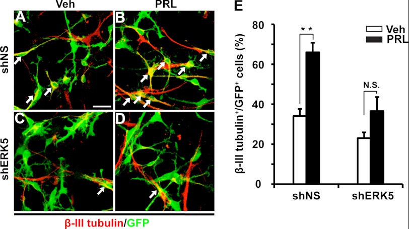
FIGURE 5.
shRNA knockdown of ERK5 decreases prolactin-induced neuronal differentiation in cultured SVZ-aNPC cells. Cells were infected with shNS (A, B) or shERK5 (C, D). Three days later, cells were treated with vehicle (Veh, A, C) or prolactin (PRL, 1 μg/ml, B, D) for 5 days. Neuronal differentiation was identified by cells expressing β-III tubulin. A–D, representative fluorescence photomicrographs of cells expressing β-III tubulin (red) and GFP (green). Arrows identify co-labeled cells. E, quantification of data from panels A–D, as the percentage of β-III tubulin+ cells in total GFP+ population. **, p < 0.01; n.s. not statistically significant. Scale bar: 25 μm.

