
FIGURE 7.
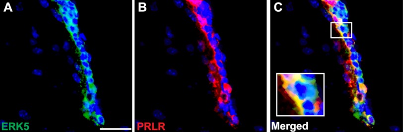
Co-localization of ERK5 and prolactin receptor in the SVZ of the adult mouse brain. Shown are representative confocal images of brain sections with Hoechst staining (blue) to identify all cell nuclei, or co-stained for ERK5 (green) (A), for prolactin receptor (PRLR, red) (B), and merged images (C). Scale bar: 25 μm.