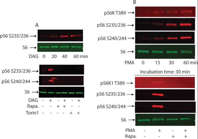
FIGURE 7.
rpS6 phosphorylation by exogenously added DAG or PMA. A, confluent and serum-starved Swiss 3T3 cells were treated with 2 μg/ml of DAG for the indicated period of time (upper panel) or treated with DAG in the absence or presence of 100 nm rapamycin or 150 nm Torin1 (lower panel). As described above, immunoblots were developed using antibodies that recognize phospho-S6 Ser-235/236, or phosphor-S6 Ser-240/244, and nonphosphorylated S6. B, serum-starved 3T3 cells were treated as above with 100 nm PMA for the indicated period of times (upper panel) or with 100 nm PMA in the presence or absence of 30 nm rapamycin for 30 min. Western blot analysis was carried out as described above.

