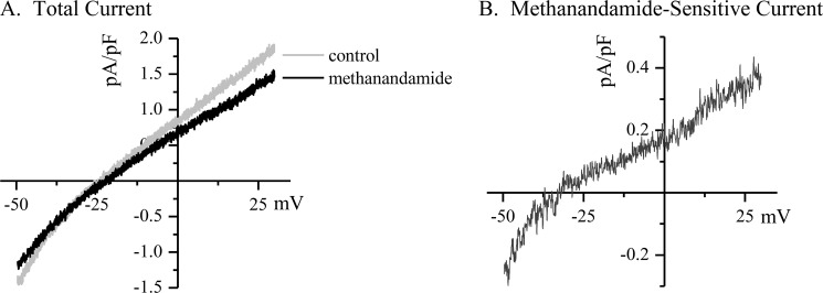
FIGURE 1.
TASK-1 is measured as the methanandamide-sensitive difference current in isolated atrial myocytes. Canine myocytes were isolated from the right atrial free wall at the time of atriotomy, and whole cell currents were measured by patch clamp recording using a ramp protocol from −50 to +30 mV. The cells were bathed in elevated external K+ (50 mm) to increase the inward component of the current and linearize the I/V curve and in the presence of CsCl (5 mm), TEA (2 mm), and nifedipine (5 μm) to block other endogenous currents. A depicts a typical trace from a canine right atrial myocyte showing the total current measured from before and after the addition of methanandamide (10 μm) to the superfusion. B depicts the methanandamide-sensitive difference current from the traces in A. The difference current is assumed to be TASK-1, and it displays the expected current-voltage relation and reversal potential consistent with a K2P channel.

