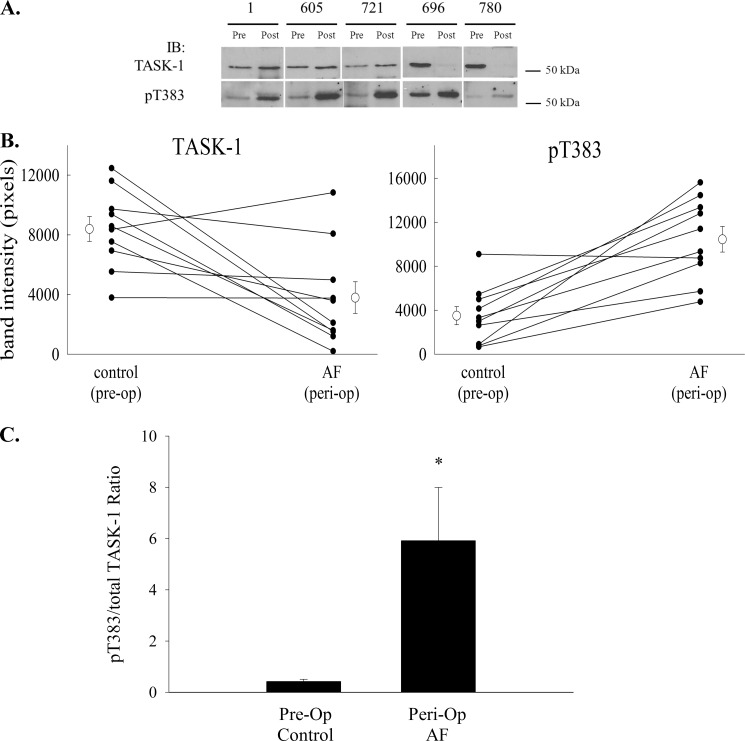
FIGURE 7.
Phosphorylation of TASK-1 at Thr-383 increases after the induction of peri-operative AF. Right atrial tissue was taken from dogs at the time of atriotomy and after induction of peri-op AF, and crude membrane fractions were prepared. Proteins were resolved by SDS-PAGE and blotted to nitrocellulose. Western blotting was used to quantify total TASK-1 and Thr(P)-383 TASK-1. A depicts several representative Western blots from tissue taken from five dogs before (Pre) or after (Post) induction of peri-op AF and immunoblotted for total TASK-1 (left panel) or Thr(P)-383 TASK-1 (right panel). IB, immunoblot. B depicts line graphs for each dog demonstrating the change in total TASK-1 (top panel) and Thr(P)-383 TASK-1 (bottom panel) before and after induction of peri-op AF. The mean (± S.E.) for all dogs is also depicted on the graph. C, relative fraction of TASK-1 phosphorylated at Thr-383 before and after peri-op AF was calculated and expressed as the ratio of Thr(P)-383 to total TASK-1. The induction of peri-op AF was associated with a significant increase in the Thr(P)-383/TASK-1 ratio (n = 10; *, p < 0.05, paired t test).

