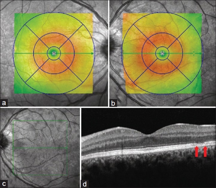
Figure 1.

a and b represent retinal thickness of the right and left eye, respectively. Perifoveal diffuse thickening is seen in the left eye. c. The infrared image reveals multiple hyporeflective lesions. d. The OCT shows discrete lesions with thinning of the photoreceptor-RPE complex (arrows)
