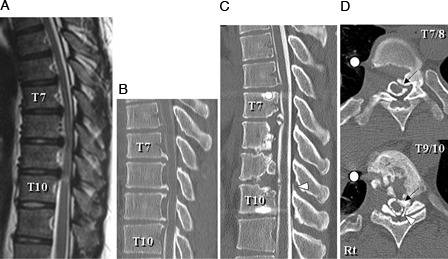
Figure 1.

Case 1: T2-weighted midsagittal magnetic resonance image (A) and CT myelogram midsagittal reconstruction plane (B) 4 years prior to this admission showing anterior compression of the spinal cord by postvertebral osseous spurs at T7–T10. CT myelogram midsagittal reconstruction plane (C) and axial planes at T7–T8 and T9–T10 (D) on admission showing re-growth of the osseous spurs that compressed the spinal cord anteriorly at T7–T8 and T9–T10 (C, D, arrows) and a newly developed ossified ligamentum flavum (OLF) that compressed the spinal cord posteriorly at T9–T10 (C, D, arrowheads).
