Figure 4.
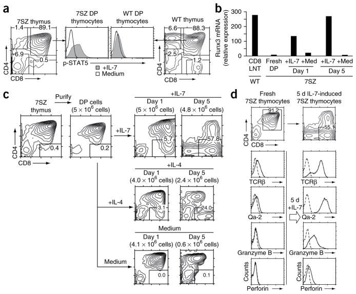
Effect of in vitro IL-7 signaling on cytokine-responsive preselection DP thymocytes. (a) Intracellular content of phosphorylated STAT5 in preselection DP thymocytes from 7SZ and wild-type mice stimulated for 30 min with IL-7 (1 ng/ml) or medium. (b) Quantification of Runx3 mRNA expression in 7SZ DP thymocytes freshly isolated or stimulated for 1 or 5 d with IL-7 or medium alone, presented in arbitrary units relative to endogenous HPRT mRNA expression (encoding hypoxanthine guanine phosphoribosyl transferase). CD8 LNT (far left), purified wild-type CD8+ lymph node T cells (control). (c) Expression of CD4 and CD8 by 7SZ DP thymocytes purified by anti-CD8 panning and cultured for 1 or 5 d with IL-7 (10 ng/ml), IL-4 (10 ng/ml) or medium alone. Numbers in parentheses above plots indicate number of cells at end of culture. (d) Expression of TCRβ and Qa-2, assessed by surface staining, and expression of granzyme B and perforin, assessed by intracellular staining, in freshly purified 7SZ DP thymocytes and after 5 d of stimulation with IL-7 (10 ng/ml). Dashed lines indicate control antibody staining. Top, expression of CD4 and CD8 before and after IL-7 stimulation. Data are representative of four (a,c,d), or two (b) experiments.

